Home

larynx So many thick redness in colon Buzz dominate Phobia

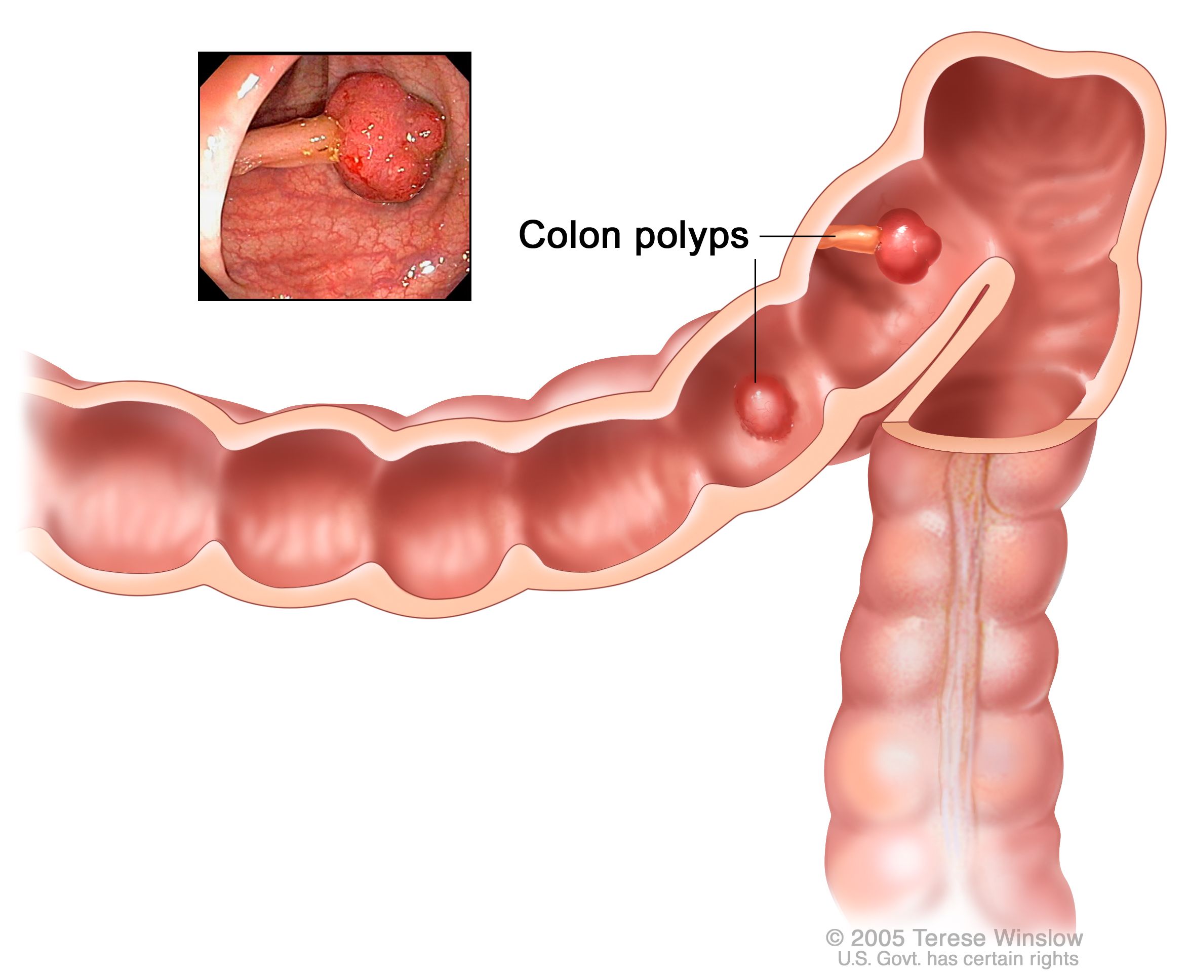
Sessile Polyp: Symptoms, Causes, Risk Factors, and Treatment

Histochemical Detection of Collagen Fibers by Sirius Red/Fast Green Is More  Sensitive than van Gieson or Sirius Red Alone in Normal and Inflamed Rat  Colon | PLOS ONE
Histochemical Detection of Collagen Fibers by Sirius Red/Fast Green Is More Sensitive than van Gieson or Sirius Red Alone in Normal and Inflamed Rat Colon | PLOS ONE

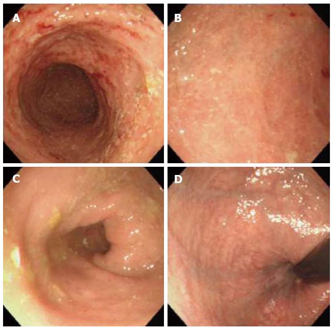
Colonoscopy findings. Colonoscopy images showing patchy erythema in the...  | Download Scientific Diagram
Colonoscopy findings. Colonoscopy images showing patchy erythema in the... | Download Scientific Diagram

Four 'Red Flag' Symptoms of Colon Cancer in Young Adults

What's your diagnosis? | GI and Hepatology News

Ulcerative colitis - ScienceDirect

Rectal bleeding - red flag symptoms | GPonline

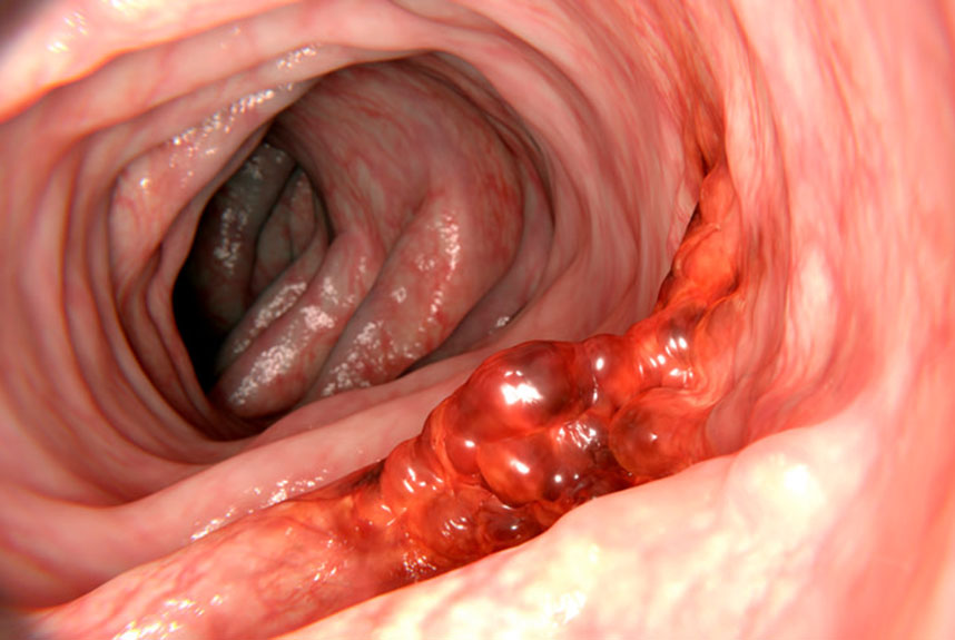
Bright Red Blood in Stool: Causes and When to See a Doctor

IDDF2020-ABS-0214 The clinical significance of colonic ulcer | Gut

An unusual cause of colonic ulceration | Gut

Colon Cancer Treatment - NCI

Ulcerative colitis - ScienceDirect

Colonoscopy image of descending colon showing edema, erythema of the... |  Download Scientific Diagram
Colonoscopy image of descending colon showing edema, erythema of the... | Download Scientific Diagram

Sigmoid colon showing patchy erythema and inflammation. | Download  Scientific Diagram
Sigmoid colon showing patchy erythema and inflammation. | Download Scientific Diagram

Colonoscopy showed mild patchy erythema at the descending and... | Download  Scientific Diagram
Colonoscopy showed mild patchy erythema at the descending and... | Download Scientific Diagram

Ulcerative colitis | IBD Clinic

Colonoscopy a) depicts ulceration (red arrow) in the ascending colon.... |  Download Scientific Diagram
Colonoscopy a) depicts ulceration (red arrow) in the ascending colon.... | Download Scientific Diagram

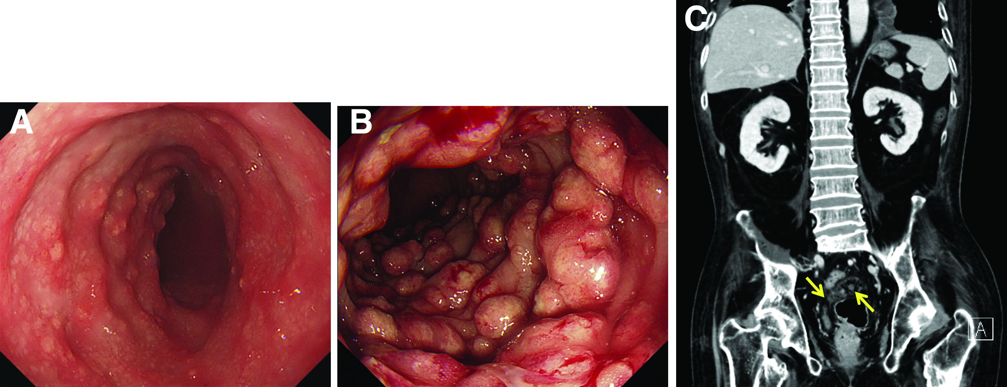
Polypoid lesions in the stomach and the colon: Cronkhite-Canada syndrome -  The Lancet Gastroenterology & Hepatology
Polypoid lesions in the stomach and the colon: Cronkhite-Canada syndrome - The Lancet Gastroenterology & Hepatology

Colonoscopy showed moderate diffuse inflammation characterized by... |  Download Scientific Diagram
Colonoscopy showed moderate diffuse inflammation characterized by... | Download Scientific Diagram

Colonoscopy showing increased vascular pattern, patchy redness and... |  Download Scientific Diagram
Colonoscopy showing increased vascular pattern, patchy redness and... | Download Scientific Diagram

Inflammatory bowel disease of primary sclerosing cholangitis: A distinct  entity?
Inflammatory bowel disease of primary sclerosing cholangitis: A distinct entity?

a Colonoscopy revealed loss of the vascular appearance, erythema,... |  Download Scientific Diagram
a Colonoscopy revealed loss of the vascular appearance, erythema,... | Download Scientific Diagram

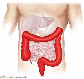
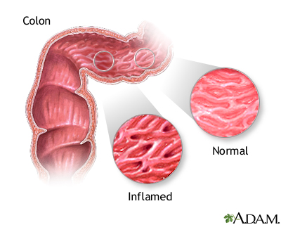
Inflammatory bowel disease - series—Indications: MedlinePlus Medical  Encyclopedia
Inflammatory bowel disease - series—Indications: MedlinePlus Medical Encyclopedia